


• Microperimetría con un amplio rango de medición.
• Prueba de fijación con un sistema de seguimiento preciso.
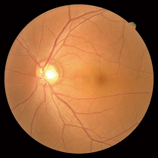
• Retinografía no midriática de alta resolución.
• Examen de retroalimentación para rehabilitación visual.
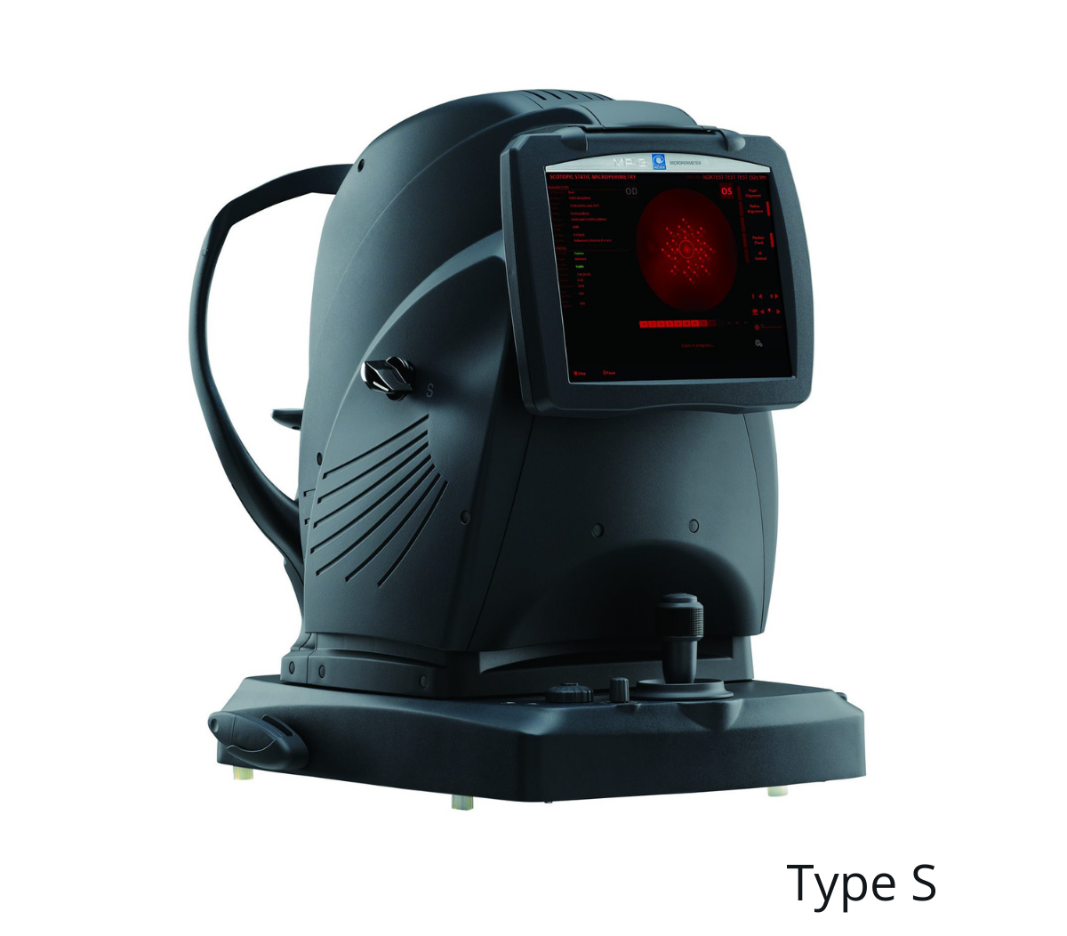
• Microperimetría escotópica
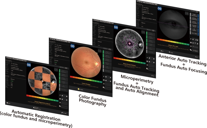
• Seguimiento automático y alineación automática.
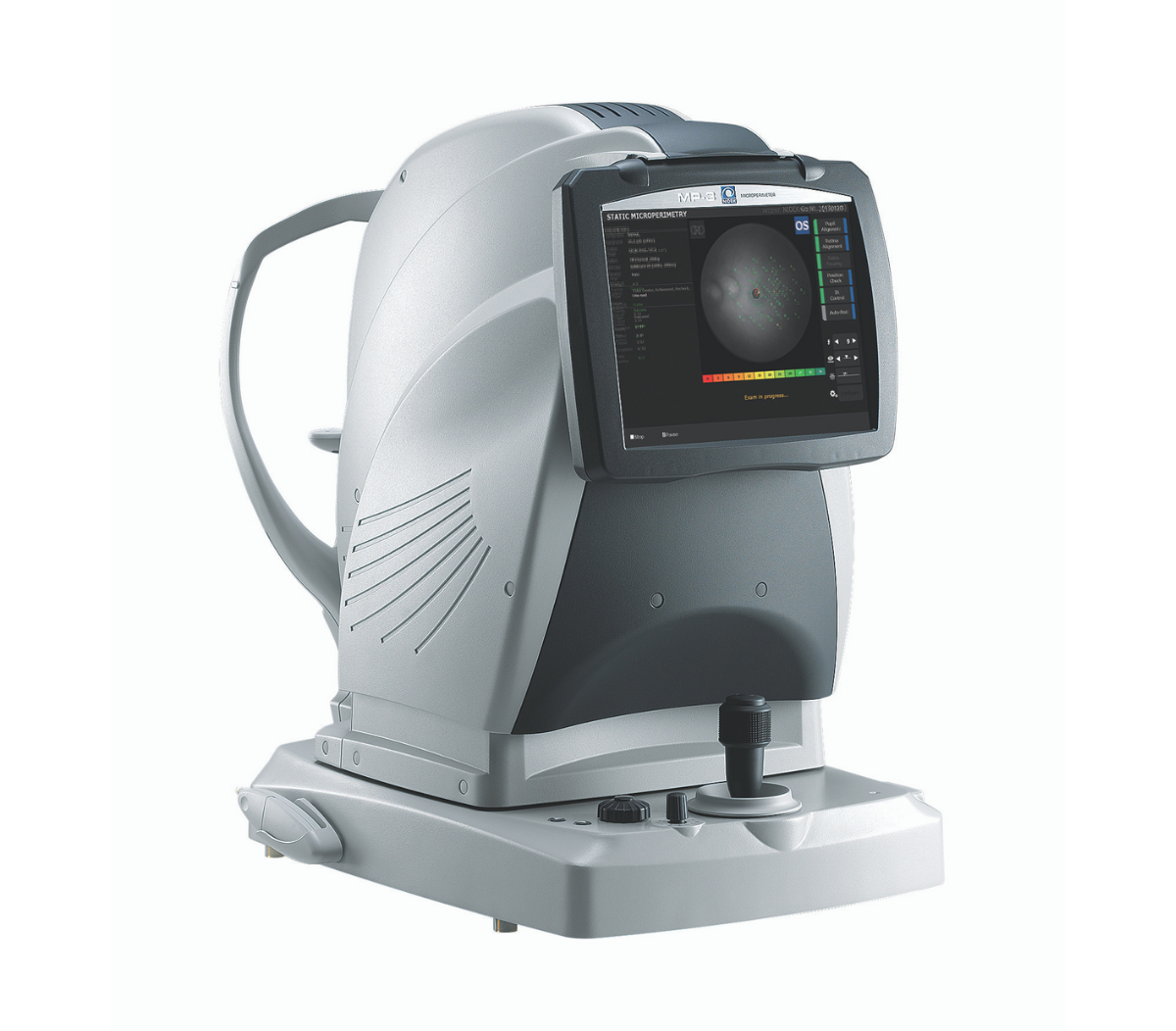
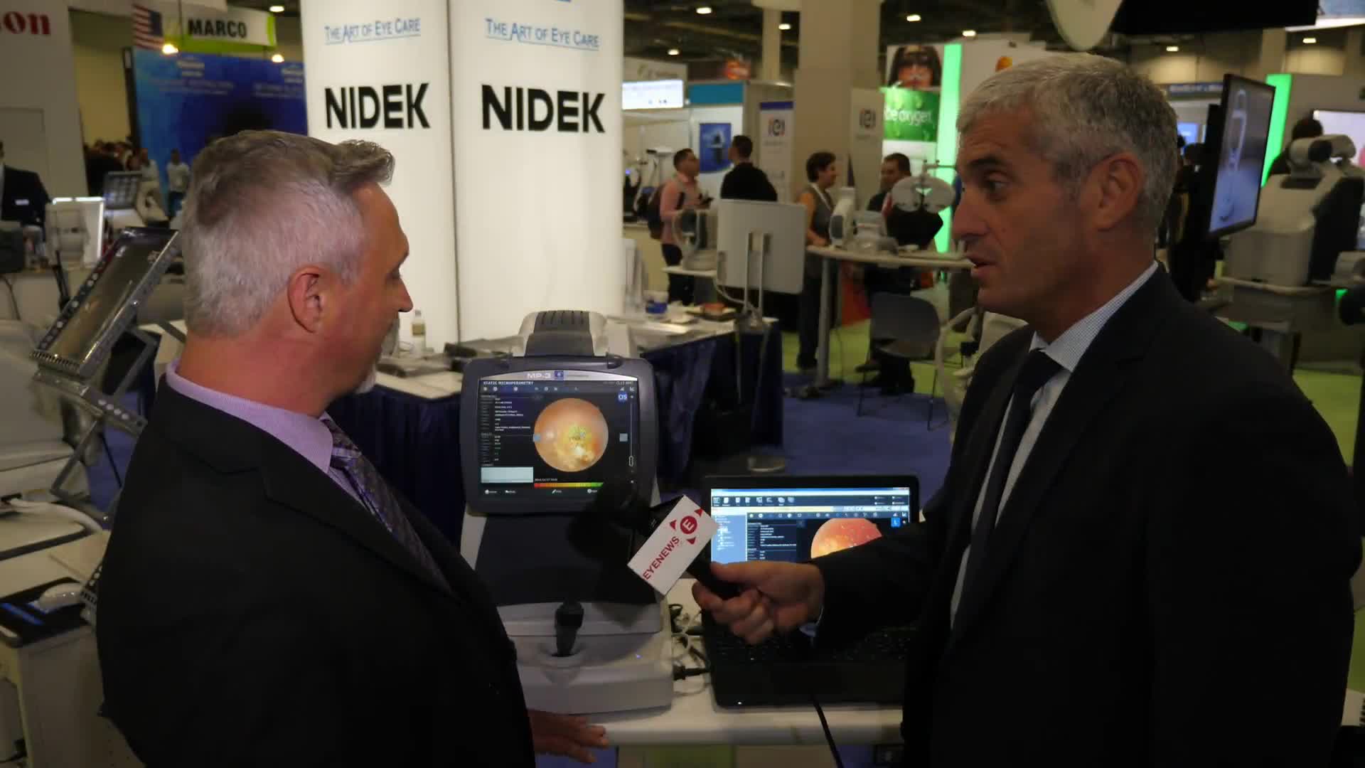
MP-3
El Microperímetro Automático
con Cámara de Fondo de Ojo No Midriática
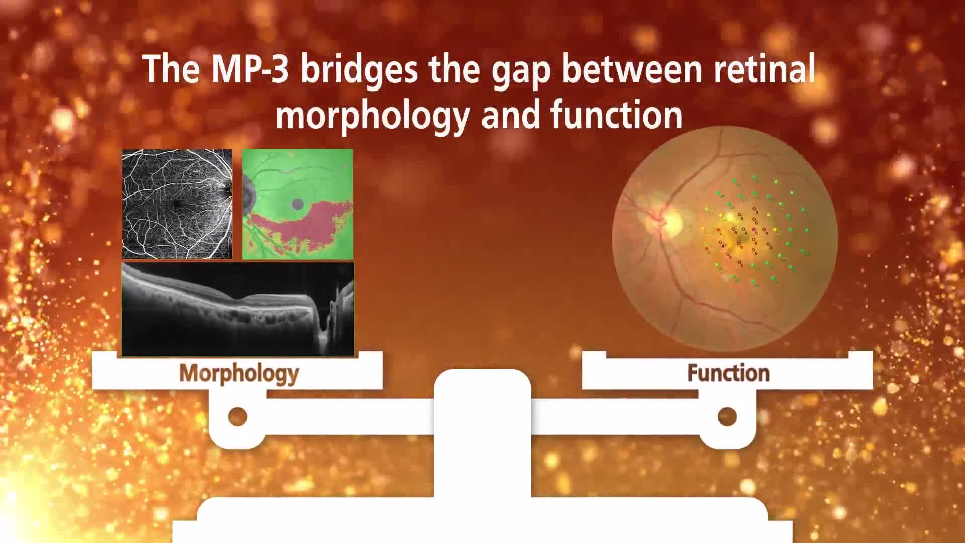
A partir de la incorporación de la tomografía de coherencia óptica (OCT, por sus siglas en inglés) en la práctica clínica, se han conseguido importantes avances en la evaluación de la morfología de retina. Además, la microperimetría dispone de una evaluación funcional avanzada de la retina.
El MP-3 mide la sensibilidad retinal local para la evaluación funcional de la retina, y utiliza los resultados para ofrecer exámenes de biorretroalimentación para entrenar a pacientes a baja visión.

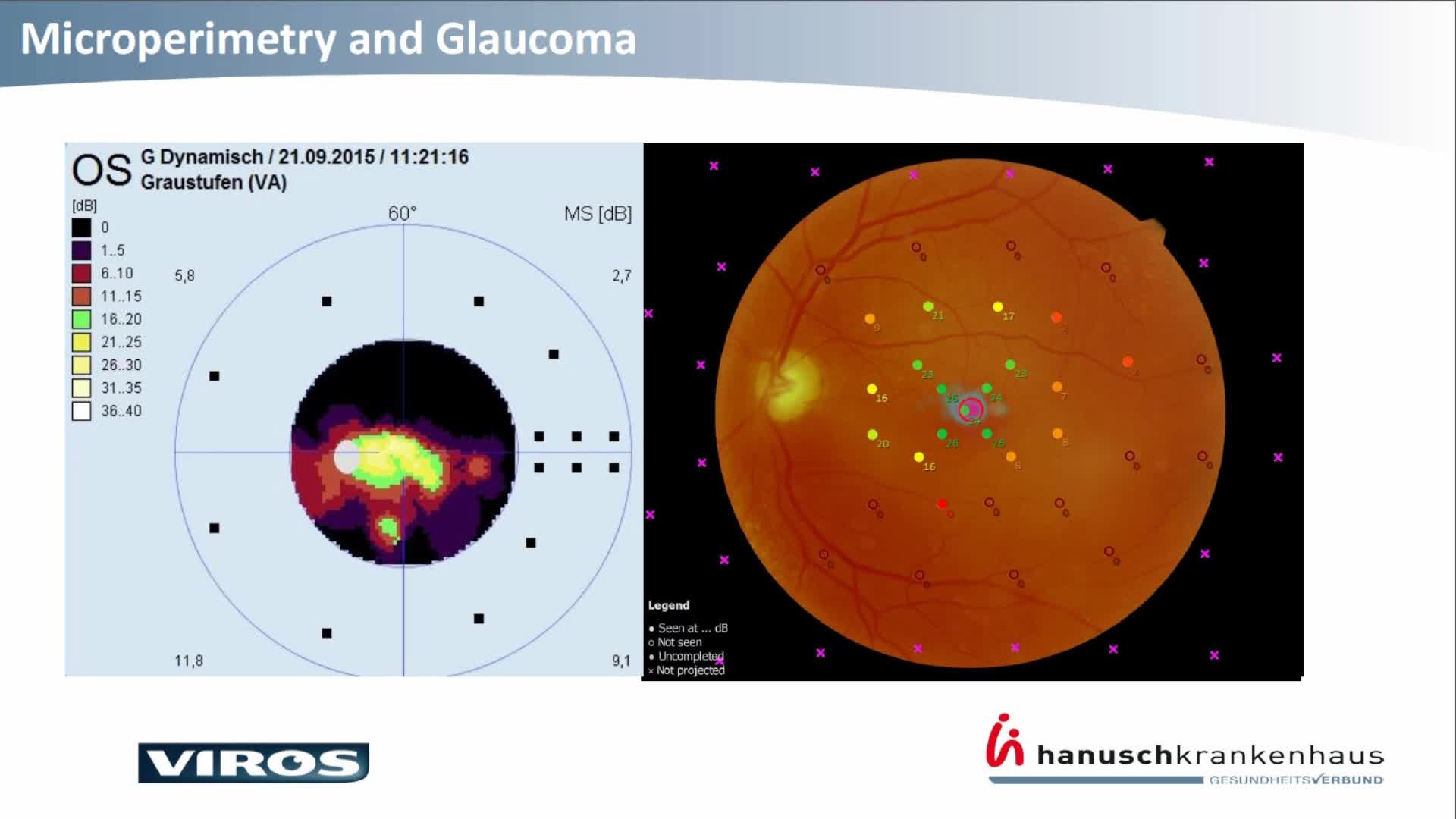
Microperimetria
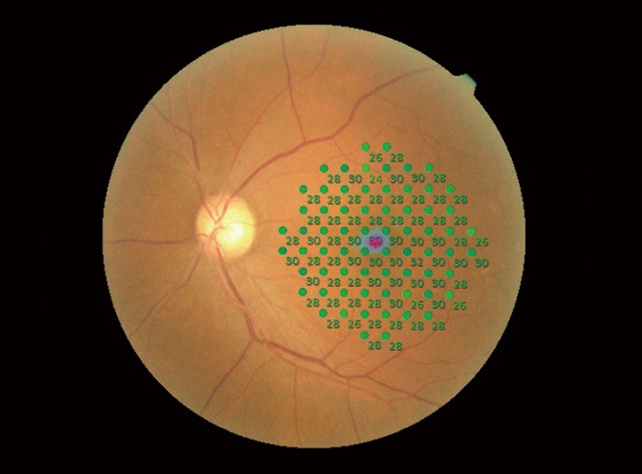
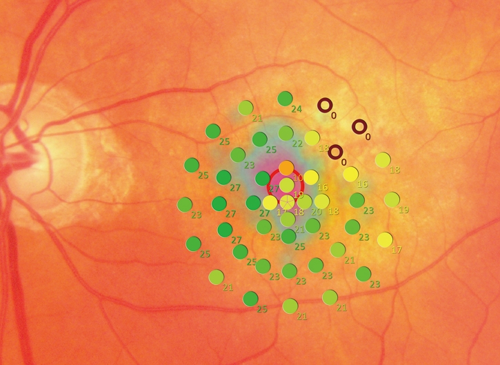
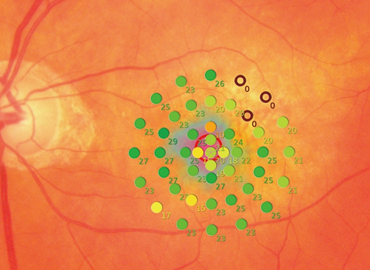
Amplio rango de medición
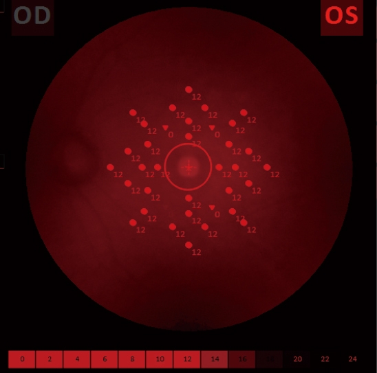
El MP-3 tiene un rango más amplio de intensidad de estímulo, de 0 a 34 dB, en comparación con el MP-1. El MP-3 mide los valores del umbral del perímetro, incluso para ojos normales. Una luminancia de estímulo máxima de 10,000 asb * permite la evaluación de baja sensibilidad.
* Según los métodos de medición ISO12866
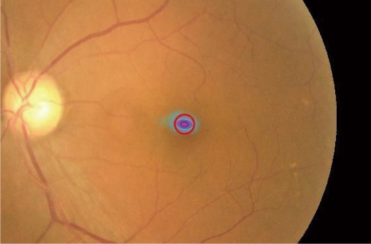
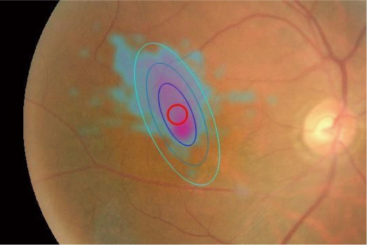
Prueba de fijación
Sistema de seguimiento preciso
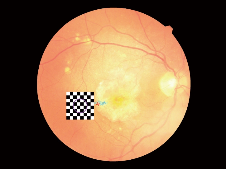
El MP-3 puede medir la fijación y determinar locus de la retina preferido, simplemente haciendo que el paciente se fije a un objetivo. El seguimiento ocular constante durante la microperimetría permite evaluar la fijación en pacientes con defectos en el campo visual central y determina si la fijación mejora después del tratamiento.
Retinografía
Retinografía no midriática de alta resolución
Una cámara de fondo de 12 megapíxeles fácil de usar está integrada en el MP-3 y adquiere imágenes de alta resolución de la patología retiniana.
Seguimiento automático y alineación automática
Las funciones de seguimiento automático y alineación automática proporcionan mediciones más precisas, lo que aumenta la comodidad y la eficiencia del paciente y del operador.
Estas funciones permiten un monitoreo sencillo y reducen las variaciones entre examinadores, lo que resulta en exámenes de seguimiento bien alineados.
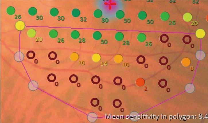
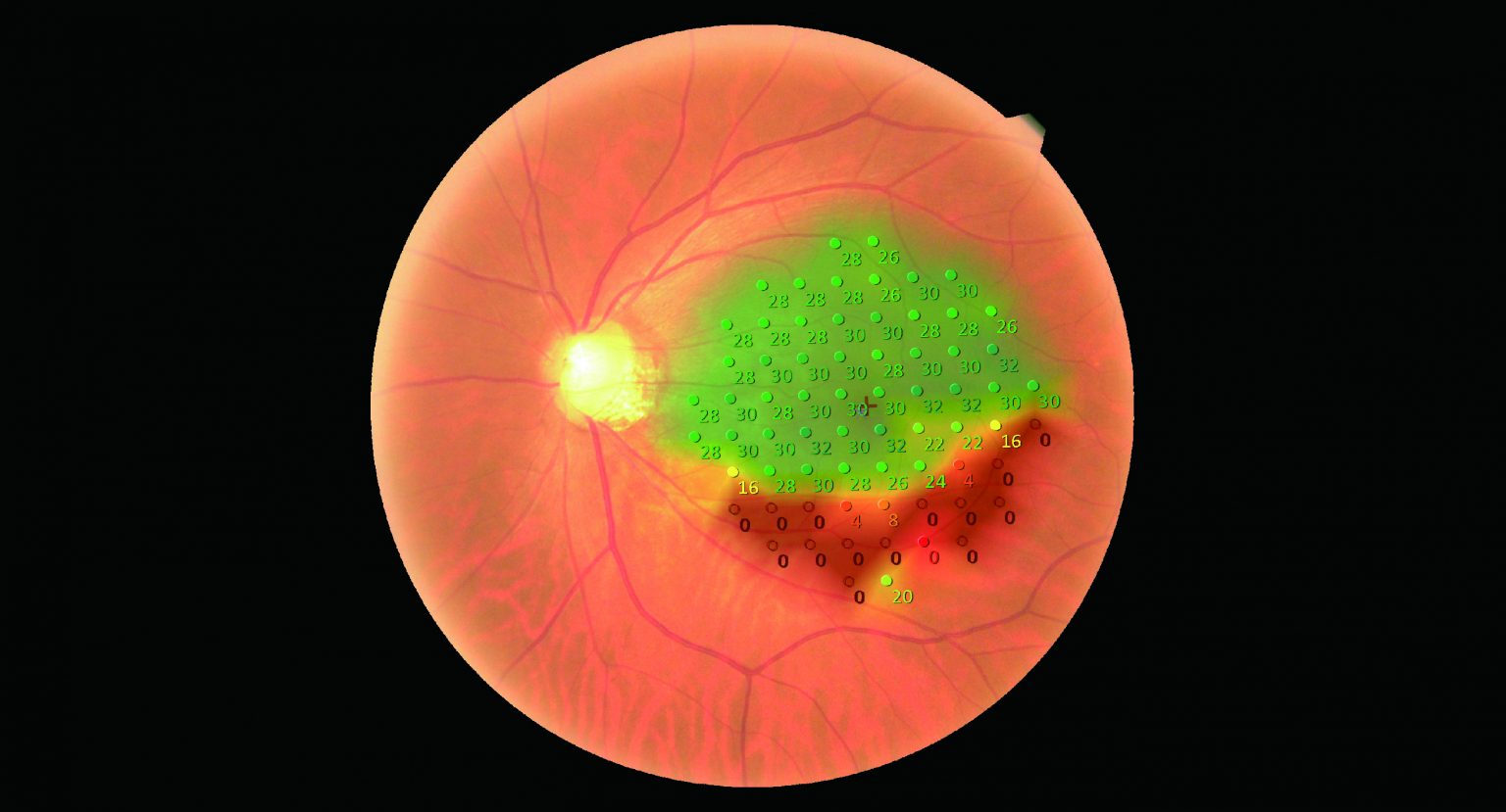
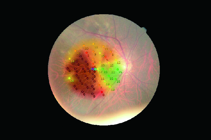
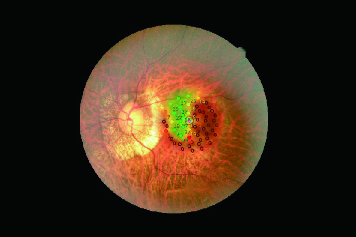
Evaluación de prueba específica de la región
Una vez completadas las mediciones, los resultados se pueden evaluar en una región específica de interés para permitir una comparación más fácil con otras imágenes de patología. Al especificar la región de interés, se muestran los resultados promedio en la región.
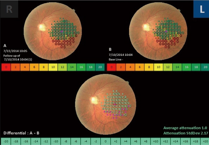
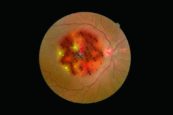
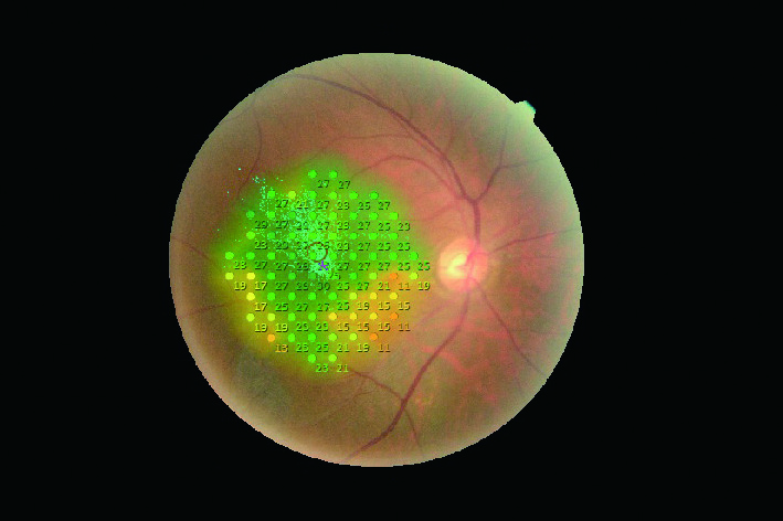
Prueba de seguimiento
Se puede realizar una prueba de seguimiento en la misma área utilizando los mismos parámetros que una prueba anterior. Esta característica le permite evaluar la progresión de la enfermedad o evaluar los resultados del tratamiento previo y posterior. Cualquier diferencia en las dos imágenes de microperimetría se muestra para una interpretación rápida e intuitiva.
Examen de Retroalimentación
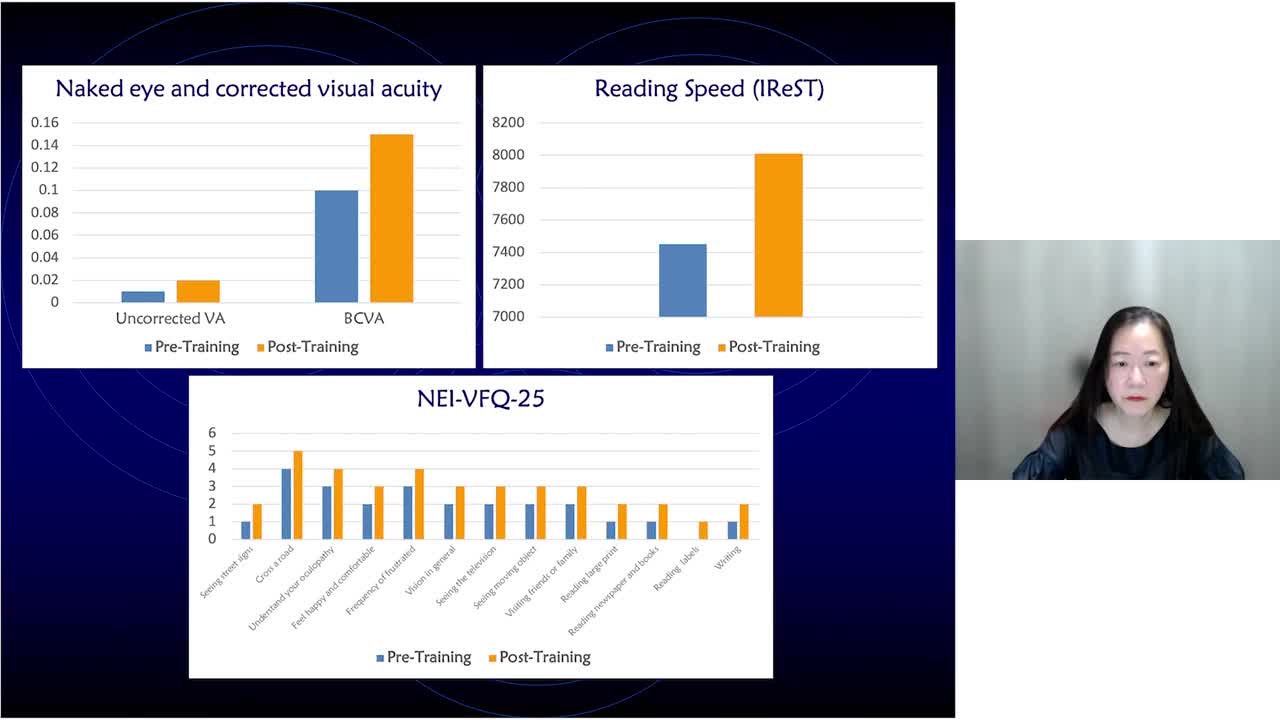
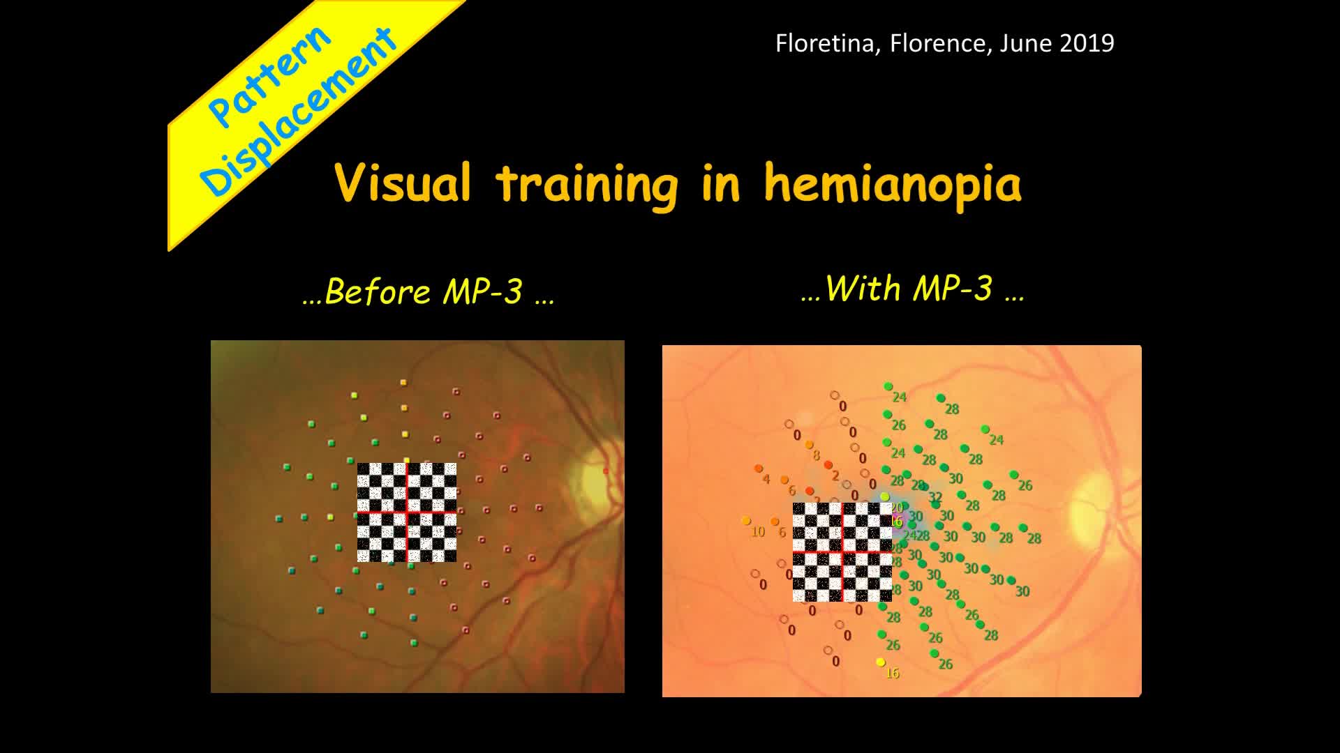
El modo de rehabilitación visual capacita a los pacientes con baja visión que han perdido la fijación foveal para reubicar su locus retiniano preferido (PRL) a una región diferente, llamada locus retiniano entrenado (TRL). El TRL está predeterminado por un médico, y la rehabilitación de la fijación le permite al paciente tener una mejor visión funcional (es decir, velocidad de lectura) debido a la mayor estabilidad de la fijación y los resultados visuales.
La estimulación activa del estándar brillante y la música alegre crean una experiencia de entrenamiento efectiva y agradable para el paciente.
Imagen cortesía del National Centre of Services and Research for the Prevention of Blindness and Rehabilitation of Visually Impaired – IAPB Italia Onlus, Roma – Italia
Artículo relacionado: Rehabilitación visual activa: un nuevo paradigma en los servicios de baja visión. Por Filippo Amore, MD, PhD
https://www.nidek-intl.com/education/case_report/retina/mp3/entry-3612.html
Microperimetría escotópica
El MP-3 tipo S mide las funciones de la retina en condiciones escotópicas (microperimetría escotópica), además de las funciones estándar del MP-3.
La microperimetría escotópica se utiliza para evaluar los cambios en la sensibilidad del bastón a las enfermedades degenerativas de la retina, incluida la degeneración macular relacionada con la edad y algunas formas de retinitis pigmentosa. Esta modalidad se puede utilizar en ensayos clínicos de nuevas terapias para enfermedades de la retina que deterioran la función del bastón
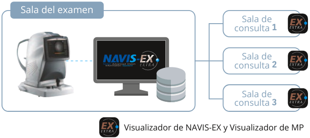
NAVIS-EX
NAVIS-EX es un software de administración de imágenes que se conecta en red con el MP-3 y otros dispositivos de imágenes de fondo de ojo NIDEK.
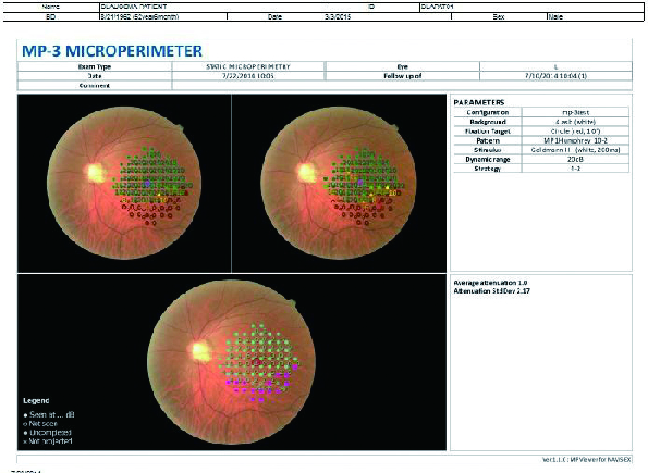
Configuración de Impresión
Hay disponibles distintos reportes impresos, incluidos los diseños especificados por el usuario cuando se utiliza con NAVIS-EX
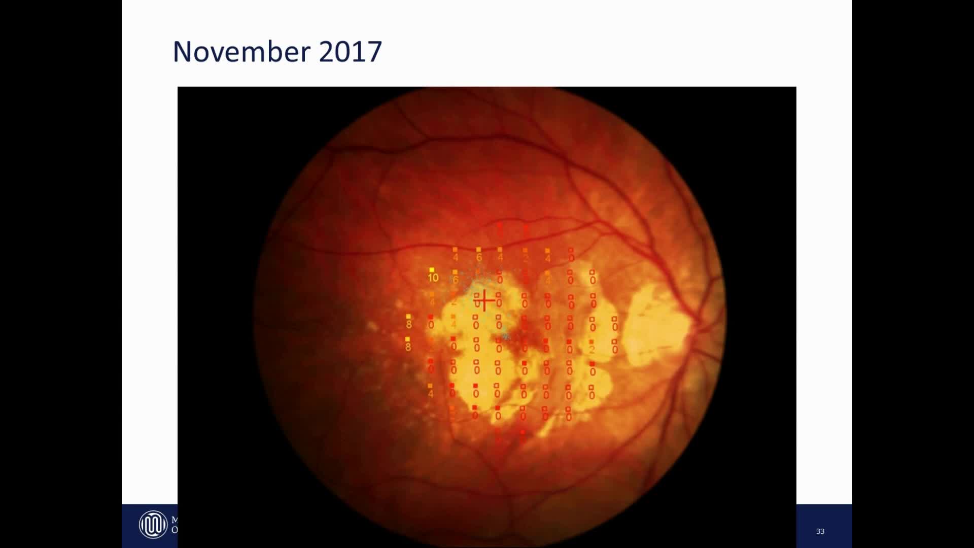
Caso de tratamiento anti-VEGF para la Degeneración Macular Relacionada con la Edad (DMRE)
Previo al Tratamiento
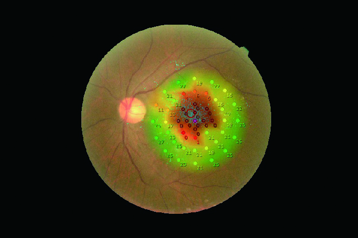
Círculo a 2° Porcentaje de puntos de fijación 66.1%
Círculo a 4° Porcentaje de puntos de fijación 92.1%
Sensibilidad media: 20,4
Posterior al Tratamiento
Círculo a 2° Porcentaje de puntos de fijación 68.1%
Círculo a 4° Porcentaje de puntos de fijación 95.5%
Sensibilidad media: 20,9
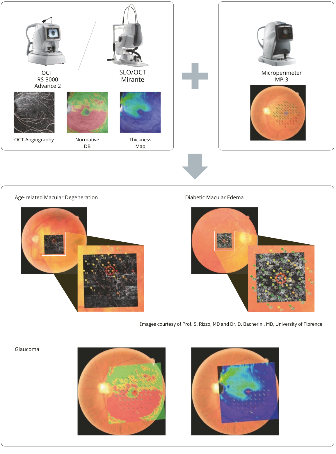
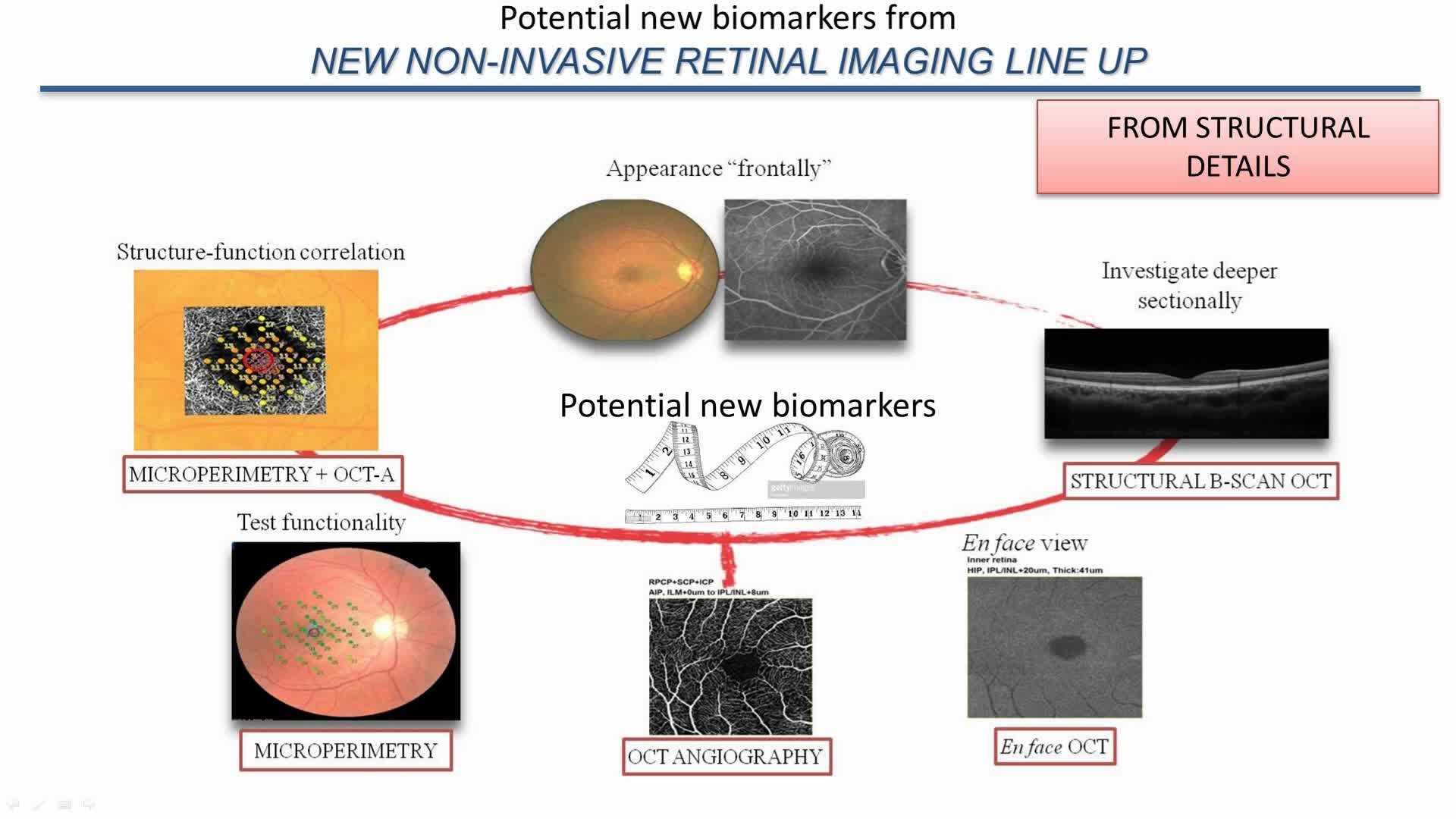
Evalúe la estructura y la función de la retina simultáneamente utilizando imágenes combinadas de OCT y microperimetría
Se pueden registrar varias modalidades de OCT capturadas por Mirante con microperimetría
Victor H. Gonzalez, MD
Gulf Coast Eye Institute, Valley Retina Institute, USA
Advances in the treatment of macular diseases will require physicians to be able to understand/measure both anatomical and functional bio markers. These measures will enable the physician to identify and follow the progress of treatment in these patients.
Microperimetry with the MP-3 has allowed me to identify patients that have loss of functional bio markers that correlate to anatomical abnormalities. For example, I can correlate loss of retinal sensitivity on Microperimetry with thickening on OCT for DME and document an improvement on retinal sensitivity with improving macular edema on OCT after anti-VEGF treatment. In this case, I treat until OCT CST and Microperimetry stabilizes. I then place the patient on treat and extend regimen until I see worsening of either Microperimetry or OCT thickening. At that point, I will continue treatment at a shorter interval until the retinal thickness normalizes and then I extend to the longest interval between injections that maintains the Microperimetry and OCT stable. A similar approach for the treatment of both dry and wet AMD will be beneficial to the patients.
Rodrigo Abreu Gonzalez, MD, PhD, FEBO
University Hospital of La Candelaria, Spain
In my clinical practice, performing microperimetry is important because it provides added value to the comprehensive evaluation of the patient. Although I don’t perform it on all my patients, when I need a comprehensive and in-depth evaluation of retinal function and structure, this is our best (and only) choice.
A practical example is our use of microperimetry to determine whether disease is active or quiescent in patients undergoing anti-VEGF treatment. Microperimetry provides the extra information that facilitates diagnostic and therapeutic decision-making. The NIDEK MP-3 provides me with ease of use, speed and total customization of the microperimetric test with the ability to integrate the structural tests with the functional outcomes using the NIDEK NAVIS-EX software.
Anna Tan, MBBS, M Med(Ophth), FRCS(Ed), FAMS
Singapore National Eye Centre, Singapore
The NIDEK MP-3 microperimetry allows me to get a detailed analysis of the macular sensitivity that can be matched to the structural imaging on OCT. This allows me to more accurately assess visual function of my patients apart from using visual acuity alone. This added detailed assessment of the macula allows me to track visual function changes in response to treatment even when visual acuity remains unchanged. The macular sensitivity representation on the microperimetry in combination with changes in OCT allows patients to clearly see how their disease is responding to treatment and helps explain to patients and their caregivers the patterns of visual losses that the patients may experience. The addition of the fixation maps allows the appreciation of the patient’s adaptation to their vision loss and is useful to plan visual rehabilitation strategies.
Sadiq N Syed, MD
Maryland Retina, USA
The NIDEK MP-3 Microperimeter has been a valuable tool in showing the patient a functional correlation to their macular condition. I will frequently perform microperimetry for patients with dry age-related macular degeneration, macular pucker, central serous retinopathy, and other macular conditions to assess whether the anatomic findings observed on examination and OCT are causing a functional deficit. In many cases significant drusen will show mild or no decrease in retinal sensitivity which is very reassuring to the patient, or if there is progressive decrease in function then you can help prepare the patient for the possibility of a future decrease in vision. The retinal tracking of this machine allows for pinpoint testing of drusen and mild RPE changes to evaluate if these physical findings are causing a functional deficit. As we are soon to have treatments for dry age-related macular degeneration, the NIDEK MP-3 Microperimeter will become an invaluable tool to show if there is treatment effect that preserves visual function in patients with this condition.
Enzo Maria Vingolo, MD
University Sapienza of Rome “Polo Pontino” Department of Sense Organs, Italy
Most of the clinical applications of Microperimetry are well known and the results supersede the most common functional tests such as Visual Acuity or Visual Field. In my practice, the NIDEK MP-3 is the optimal device for visual rehabilitation. We use this device to understand the functionality of the residual retinal sensitivity, modifications to the cloud of fixation, and to localize the Preferred Retinal Location (PRL) that often occurs after clinical damage.
If the combined outcomes of these tests indicate decreased functional vision, then we can use the Biofeedback training available in MP-3 to change the PRL location.
A Training Retinal Location (TRL) allows better visual performance for reading and common daily tasks.
Additionally, this strategy frequently results in decreasing the magnification power required by the patient, resulting in use of more friendly visual aids.
The latest use of this application is to improve performance among athletes. Training the normal fovea to increase speed of fixation saccades is very useful in sports involving fast moving targets or team players (eg. volleyball, shooting, race-car drivers, etc.).
Thanapong Somkijrungroj, MD
Chief of Uveitis Unit, Retina Unit, King Chulalongkorn Memorial Hospital and University, Thailand
Structural assessment with multimodal imaging is becoming the standard in diagnosis and management of acquired and hereditary retinal diseases. In contrast, visual function is generally evaluated by visual acuity and traditional visual field (VF) tests which have well-documented limitations. This is especially important in patients with retinal pathology and compromised fixation that impacts the reliability of VF testing.
Retinal sensitivity evaluated by microperimetry bridges the gap between functional and structural evaluation in retina diseases. Additional benefits of microperimetry include the identification of the preferred retinal locus (PRL), low vision training and to serve as an outcome measure in clinical trials.
Hence, in addition to structural assessment with multimodal imaging, microperimetry empowers physicians to assess visual function beyond the limits of visual acuity and conventional perimetry.
Laurentino Biccas Neto, MD, PhD
Ocular Vitória, Brazil
Fundus-based perimetry is an essential tool for the comprehensive retinal specialist. It allows an objective assessment of retinal function and a better understanding of macular physiology. This is also very relevant in decision-making, using thresholds for treatment indication, such as early photodynamic therapy for low-sensitivity-central-serous retinopathy, and for determining prognosis during preoperative evaluation.
It may prove to be a very valuable resource in diagnosing poorly visible inflammatory lesions, such as some white dot syndromes, and in following up patients with distorted macular architecture and new-onset complaints, such as macular dragging in FEVR. Assessment of central fixation and biofeedback stimulation is also a new and fascinating utility to be explored in our low vision patients.
Thales A. C. de Guimarães, MD
Moorfields Eye Hospital NHS Foundation Trust, UK
UCL Institute of Ophthalmology, UK
The MP-3 Microperimeter is a versatile and powerful platform to study retinal sensitivity in individuals affected with sight-threatening disorders. More often than not, diseases of the retina display a disconnection between structure and function, which is particularly noticeable in individuals with inherited retinal disorders. I have been impressed with the performance of the MP-3 even under the most difficult circumstances, such as patients with nystagmus and eccentric fixation. With the faster tracking speed coupled with the high-resolution fundus camera, ease-of-use, and the ability to quickly customize each test individually, this microperimeter has been a game changer. It has enhanced my clinical understanding and perspectives of certain retinal diseases, providing new research opportunities – all to benefit patients. In the era of personalized medicine, the MP-3 is a one-of-a-kind tool that allows for more adequate disease prognostication and follow-up.