
- A melhor plataforma de captura de imagem multimodal
Mirante SLO/OCT:
-
- Modo Colorido/ FA /ICG /FAF-Azul/ FAF-Verde/ Retro
- OCT/ Angiografia-OCT*
Mirante SLO:
-
- Modo Colorido/ FA*/ ICG*/ FAF-Azul/ FAF-Verde/ Retro
- Campo ultra amplo x imagem ultra HD
- Cor insuperável
- FA e ICG dinâmicos/ simultâneos
- Modo Retro exclusivo
- OCT de área ampla em HD
- Função Fly Through
* Opcional
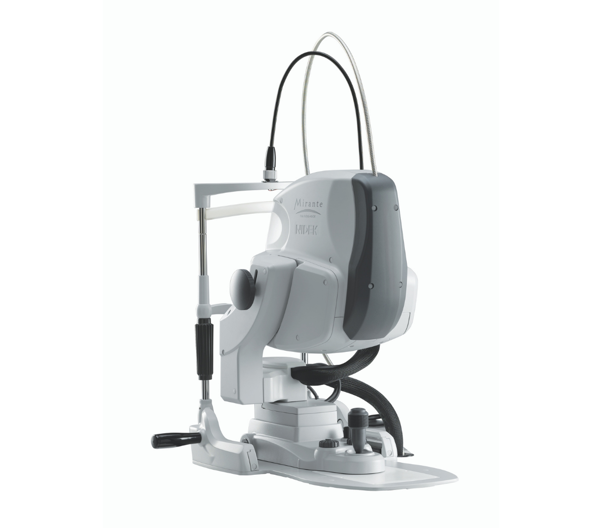
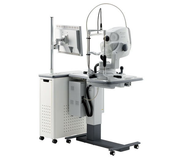
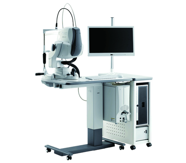
Mirante SLO/OCT
Mirante SLO
A Melhor Plataforma de Imagem Multimodal
Combinação de SLO & OCT de Última Geração
Imagem Ultra Wide Field x Ultra HD
Uma combinação impressionante de Campo Ultra Amplo de 163° x HD Ultra 4K incorporada no Mirante proporciona uma visão mais ampla e aprimorada da estrutura da retina e da vasculatura com clareza incomparável.
")
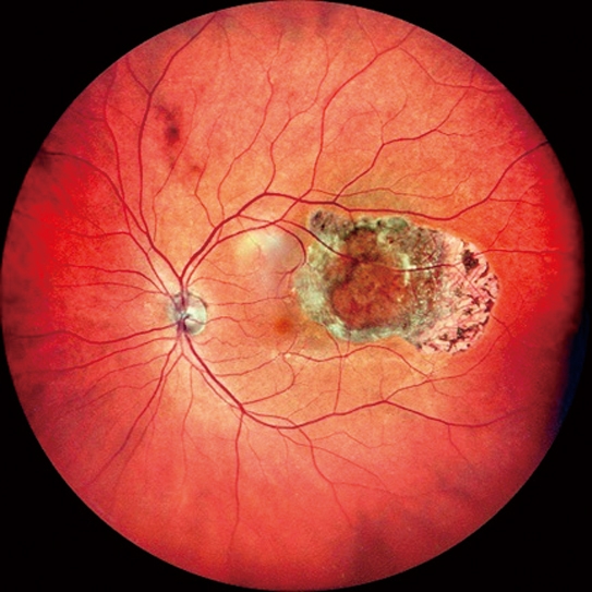
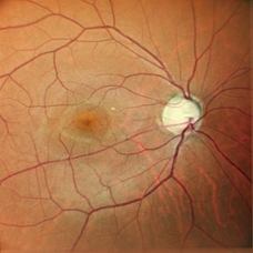
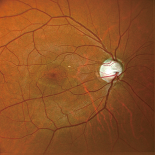
SLO colorido
Cor e clareza incomparáveis para cada detalhe.
-
-
- Imagem colorida de campo ultra amplo de 163°
- Composição de imagem panorâmica
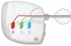
- Detectores triplos RGB
- Cor RGB + exibição de cores selecionáveis com uma única captura
-
FlexTrack
O novo algoritmo FlexTrack corrige distorção de imagem devido à fixação instável e melhora a qualidade da média.
Modo Retro / FAF
Modalidades não invasivas de valor agregado expandindo sua prática.
-
-
- Modo Retro
- FAF-Azul/ FAF-Verde
(autofluorescência do fundus)
-
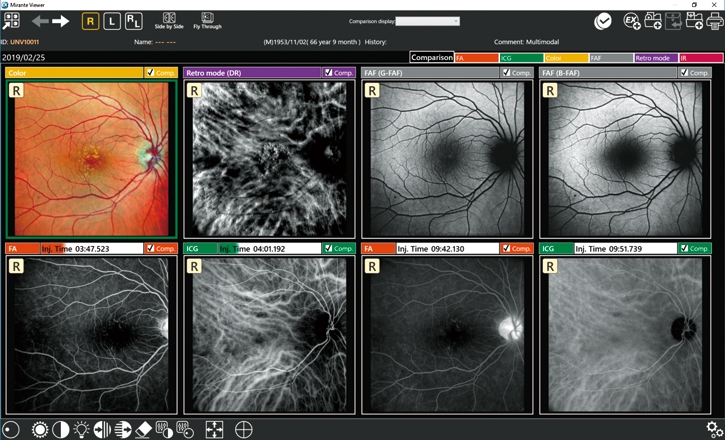
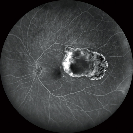
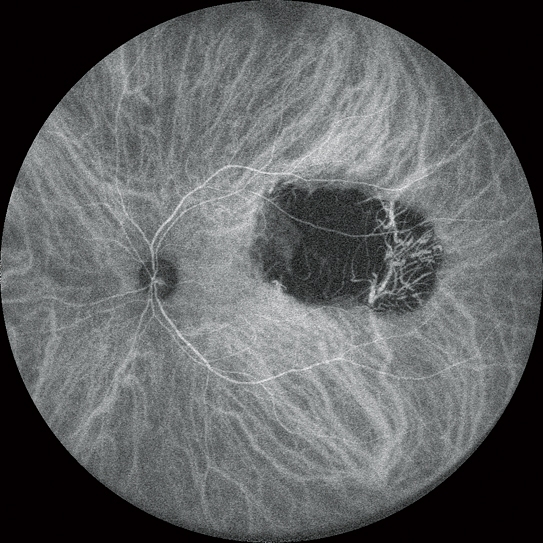
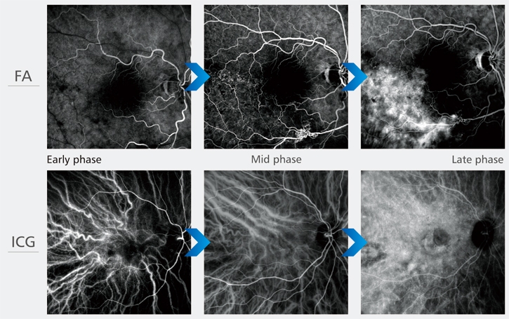
FA e ICG
Avaliação eficaz do fluxo sanguíneo dinâmico e estático.
-
-
- Imagens FA e ICG de campo ultra amplo de 163°
- Angiograma dinâmico e estático em HD
- FA e ICG simultâneos
- Fácil comparação de FA e ICG
-

OCT
Captura de imagem em HD com uma variedade de funções
-
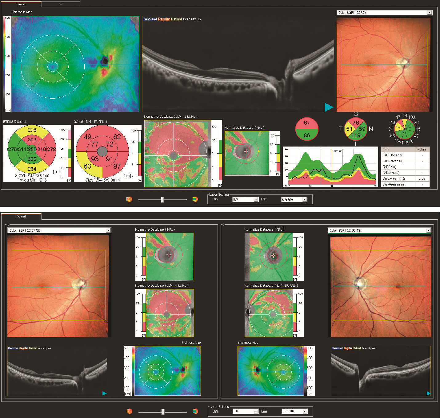
- OCT de área ampla em HD
- Análise de glaucoma
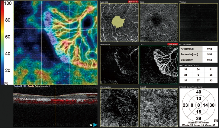
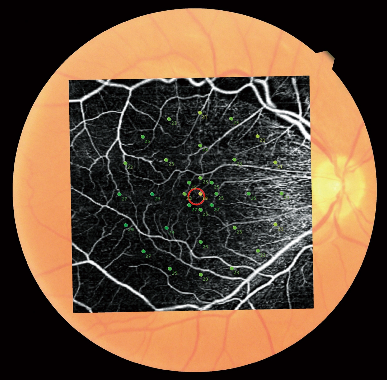
AngioScan
-
- Mapa de densidade de vasos e mapa de densidade de perfusão
- Autodetecção de FAZ e análise de forma
- Varredura de área ampla
- Tracing HD plus
- Definição selecionável

Apresentação do Produto: Mirante SLO/OCT
Demonstração do Produto: Mirante SLO/OCT
Demonstração do Produto: Mirante Viewer / NAVIS-EX
Message from Senior Product Specialist
Message from the Engineers
Para o modelo Mirante SLO/OCT:
Modo Colorido/ FA/ ICG/ FAF-Azul/ FAF-Verde/ Retro
OCT/ Angiografia-OCT*
Para o modelo Mirante SLO:
Modo Colorido/ FA*/ ICG*/ FAF-Azul/ FAF-Verde/ Retro
*Opcional
Captura de imagem de combinação simplificada
A captura de imagem Combo permite a captura sequencial de imagens com a combinação predefinida de configurações de captura de imagem para cada doença especificada.
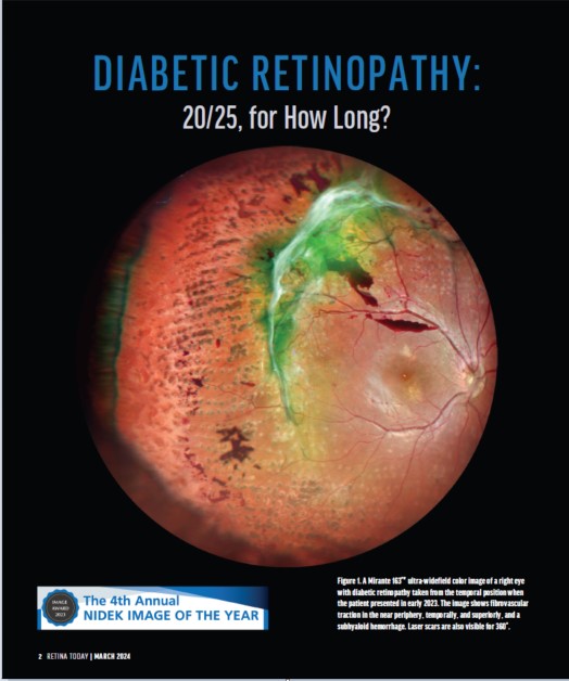
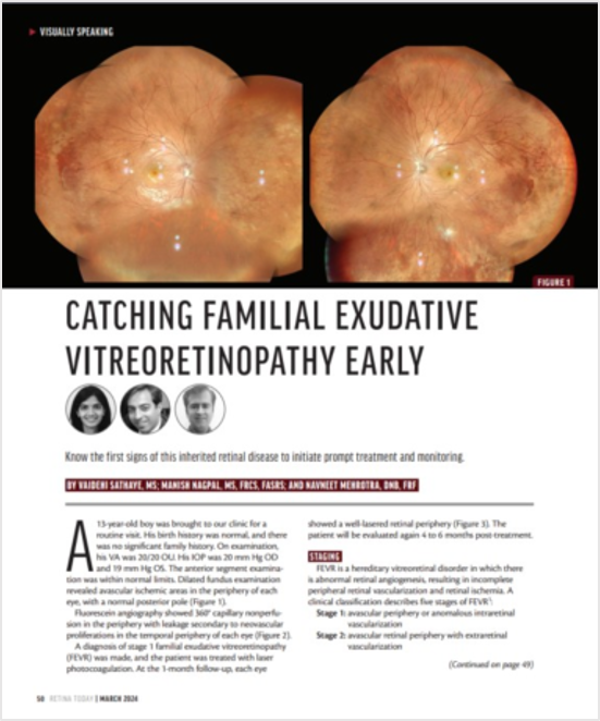
Imagem de Campo Ultra Amplo de 163°
A imagem nítida de todo o campo de visão de 163° permite uma avaliação detalhada das patologias da fóvea à periferia extrema.
(Imagens de campo ultra amplo estão disponíveis com o adaptador de campo amplo opcional).
Ultra 4K HD e Função Média para Clareza Incomparável
As imagens de 4.096 x 4.096 pixels capturam todos os detalhes da retina e da coroide. Além disso, o zoom permite alta ampliação, visualização clara de mudanças sutis na patologia e resolução dos detalhes finos dos capilares.
O algoritmo FlexTrack corrige a distorção da imagem devido à fixação instável e melhora a qualidade da média.
Três detectores RGB separados varrem simultaneamente diferentes profundidades da retina com comprimentos de onda vermelho, verde e azul. Um histograma em cores está disponível para ajuste fino com base na patologia ou na preferência do profissional.
Angiograma Dinâmico em HD
Os vídeos podem ser gravados com no máximo 1.024 x 1.024 pixels por até 120 segundos. Vários vídeos curtos podem ser gravados durante a mesma medição.
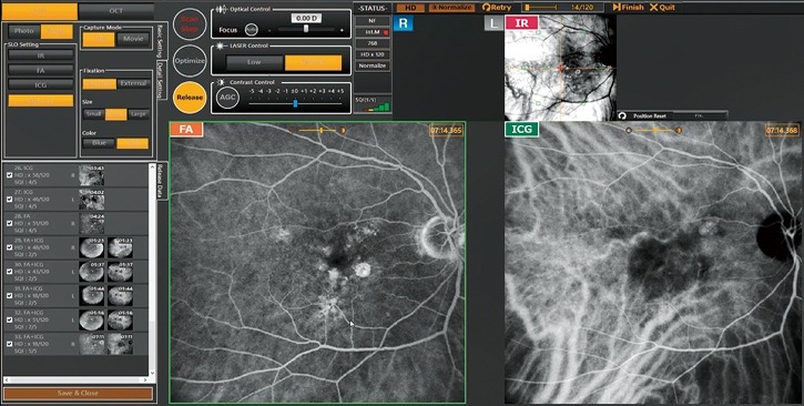
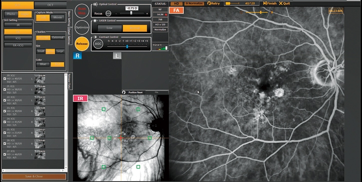
FA e ICG Simultâneos
O Mirante permite a obtenção simples e simultânea de imagens de FA e ICG.
O monitoramento IR ao vivo permite o alinhamento antes da emissão de fluorescência e reduz o risco de perder uma fase muito precoce da angiografia.
O controle automático de ganho (AGC) ajusta simultaneamente o contraste de cada imagem de FA e ICG, tornando a captura de imagem do fluxo sanguíneo dinâmico um procedimento muito simples.
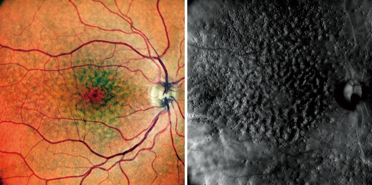
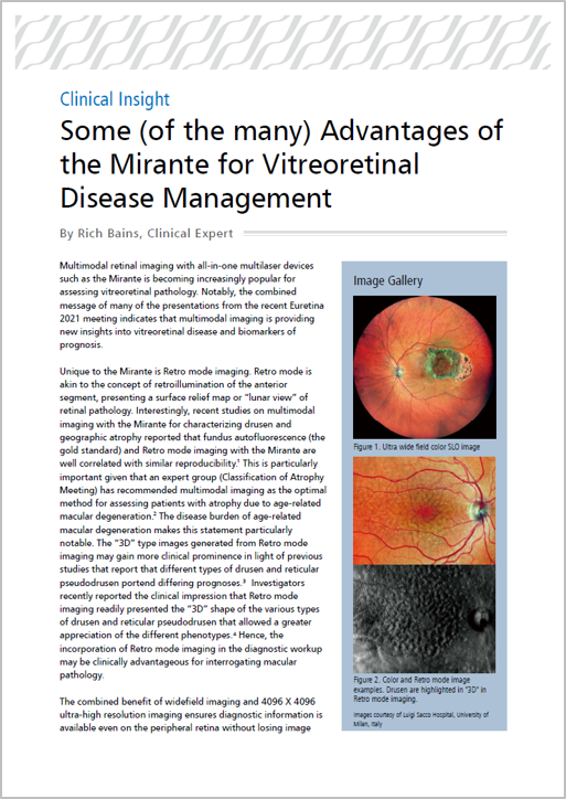
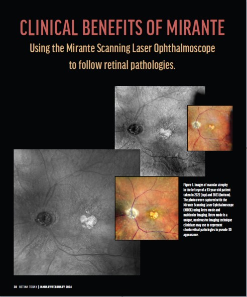
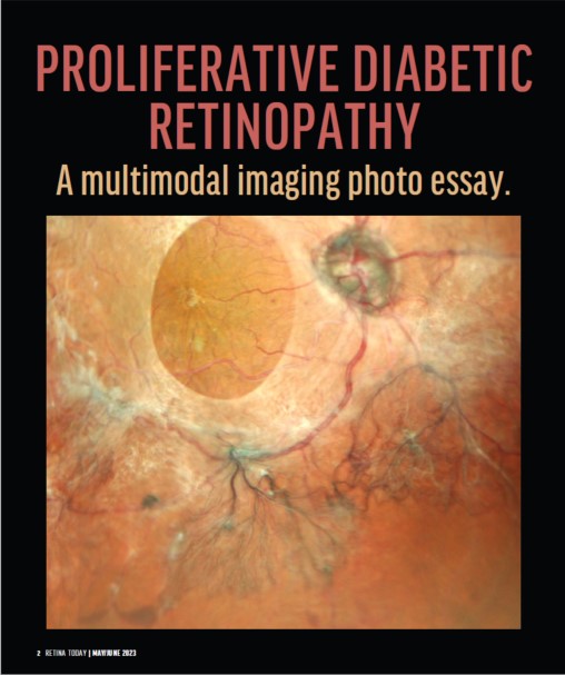
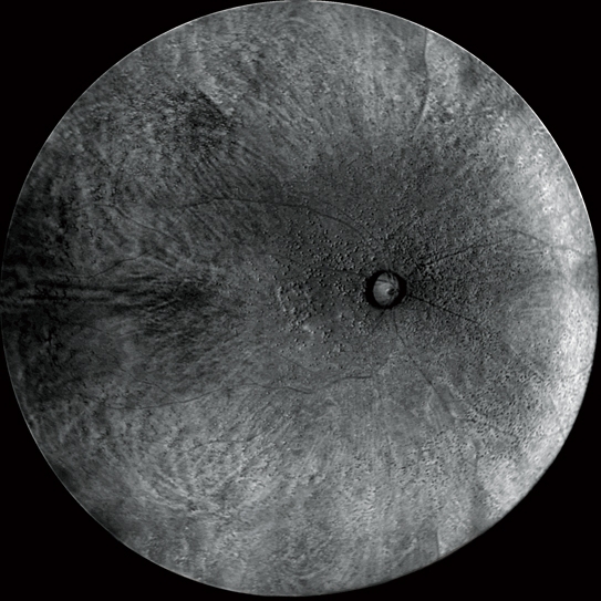
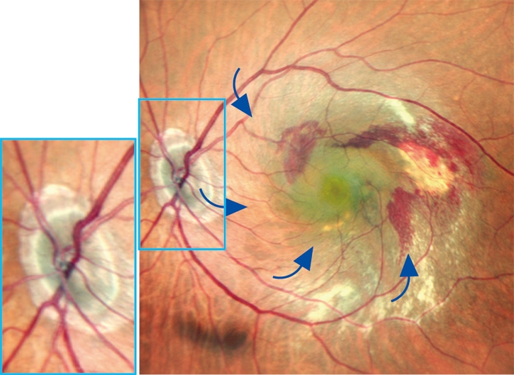
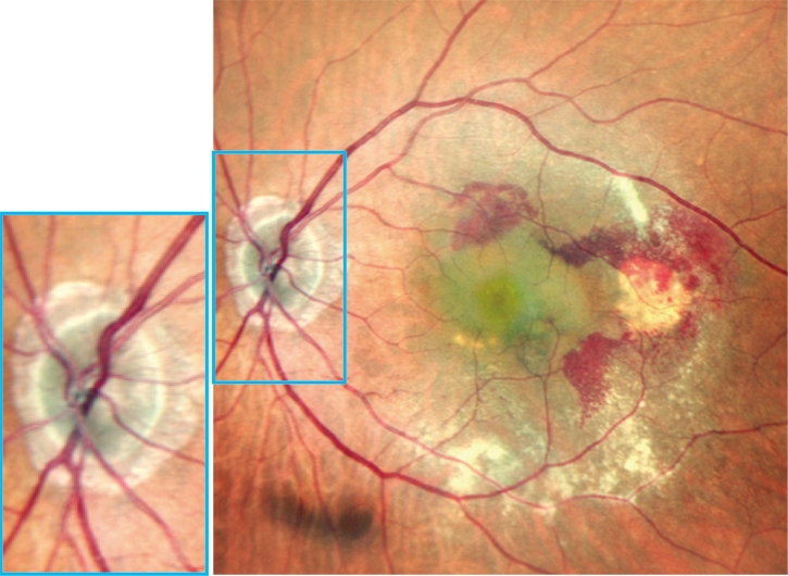
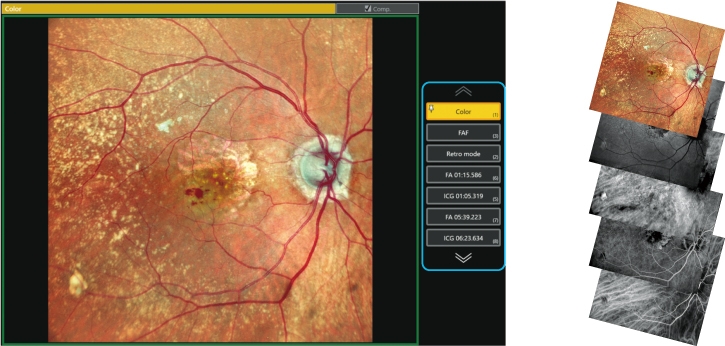
O modo retro é uma técnica não invasiva exclusiva para detectar alterações patológicas na coróide.
Essa modalidade de captura de imagem usa luz infravermelha dispersa para detectar reflexos anormais na coróide causados por drusen, edema e outras patologias coriorretinianas sutis.
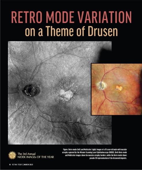
Imagens em modo colorido e retrô (Drusen)
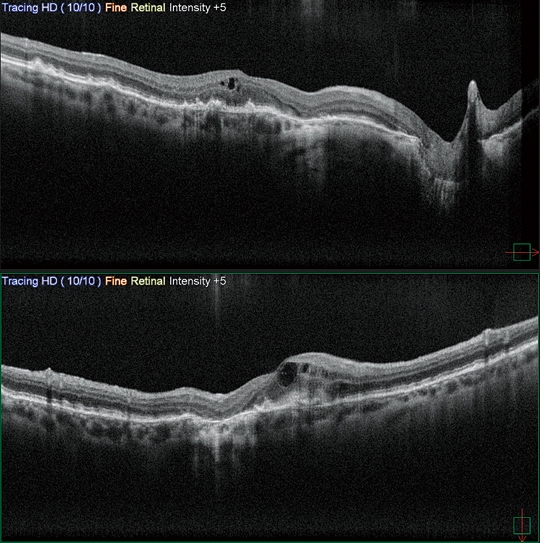
O Retina map permite o diagnóstico de área ampla, incluindo a mácula e o disco óptico em um único disparo. O modo ultrafino e as funções de rastreamento em HD plus fornecem imagens de alta qualidade para observação detalhada do vítreo ao coróide.
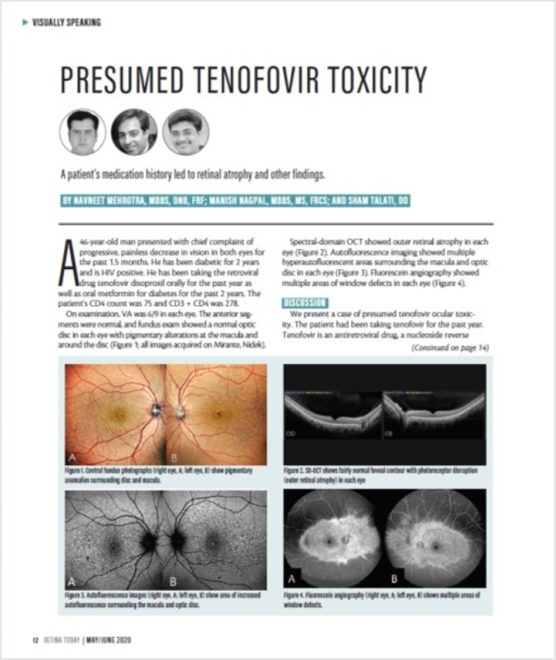
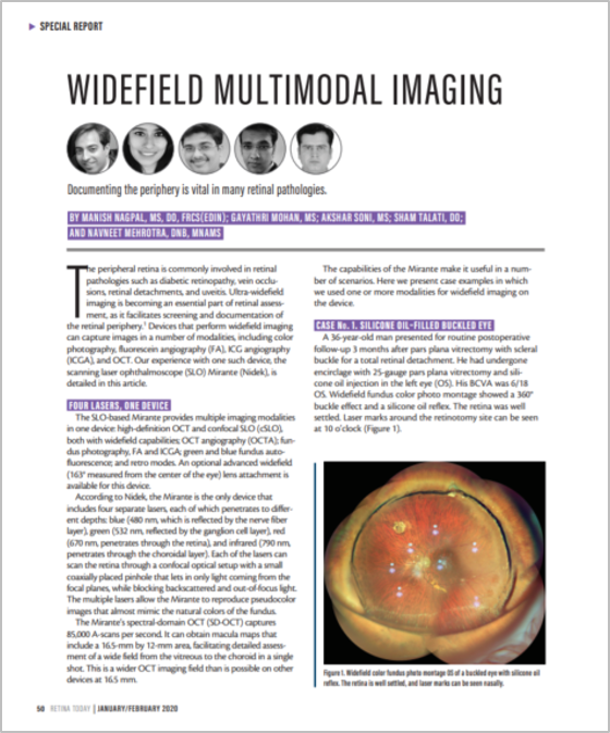
*Imagens cortesia do Hospital Luigi Sacco, Universidade de Milão, Itália/ Fundação Retina e Centro de Pesquisa Oftalmológica, Índia
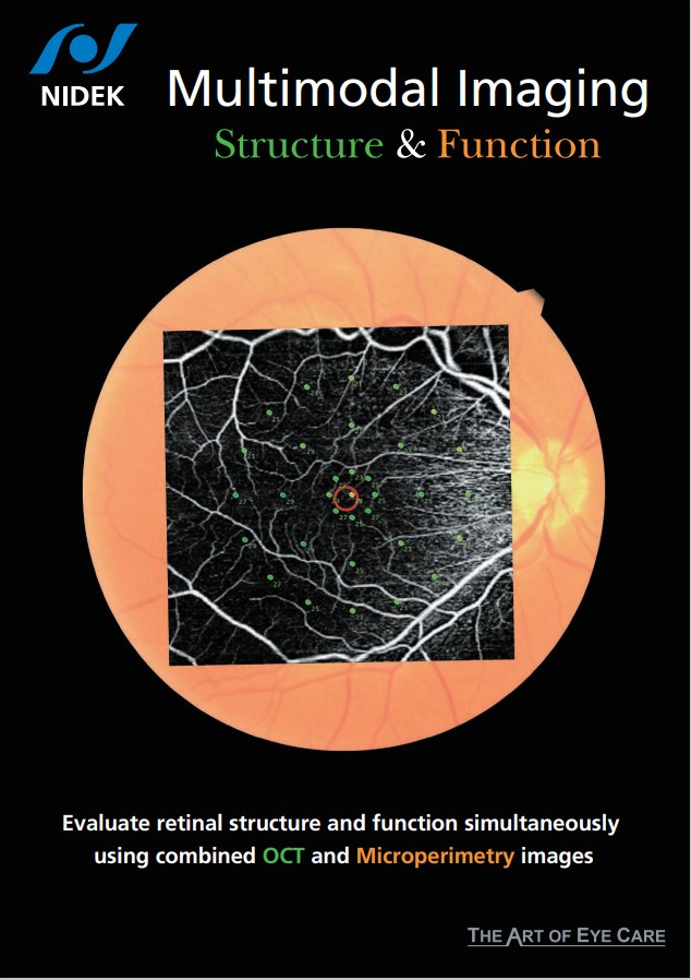
A função Fly Through aprimora ainda mais a imagem multimodal, registrando e sincronizando imagens de diferentes modalidades para visualizar a mesma área enquanto percorre a região de interesse.
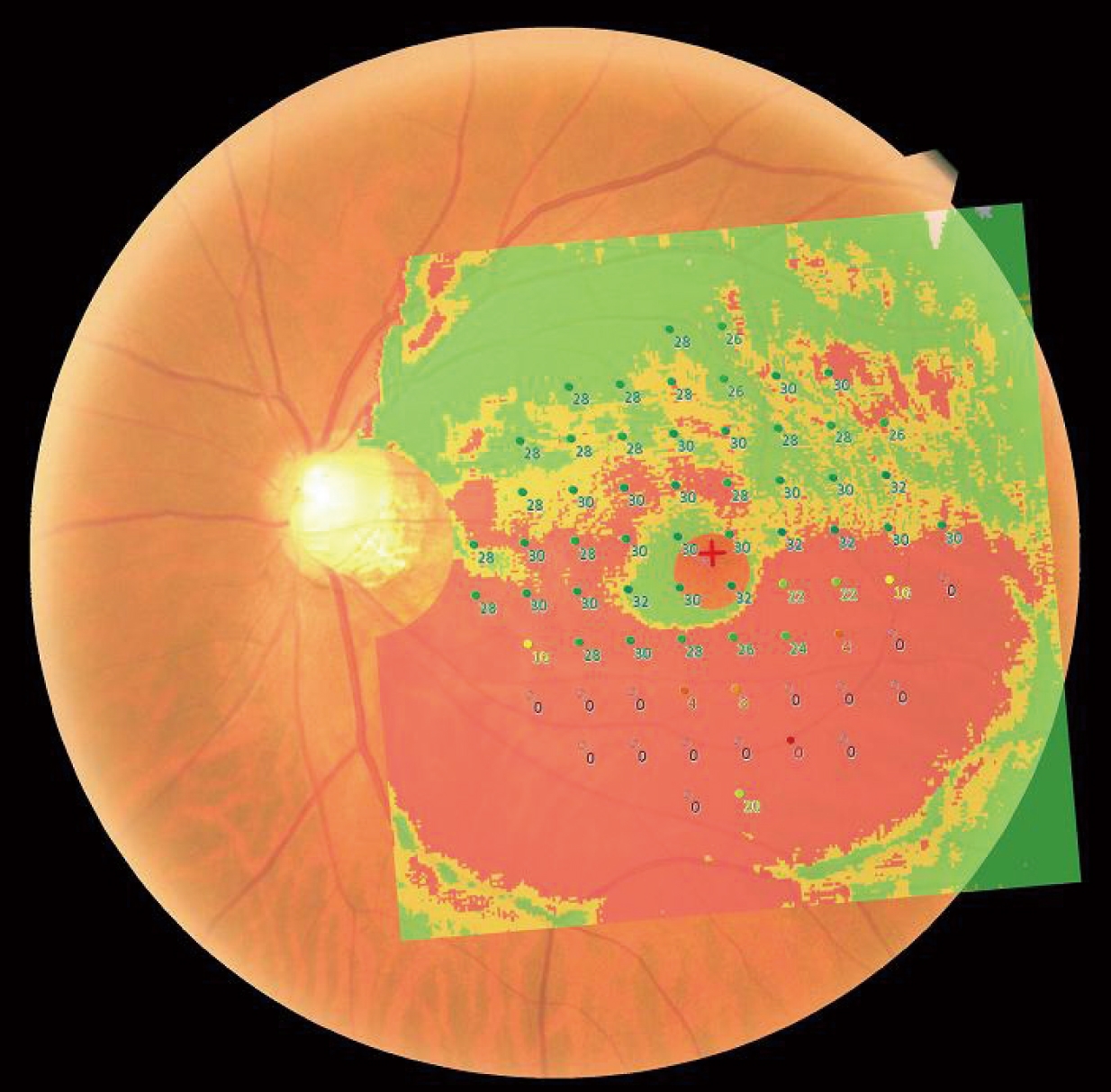
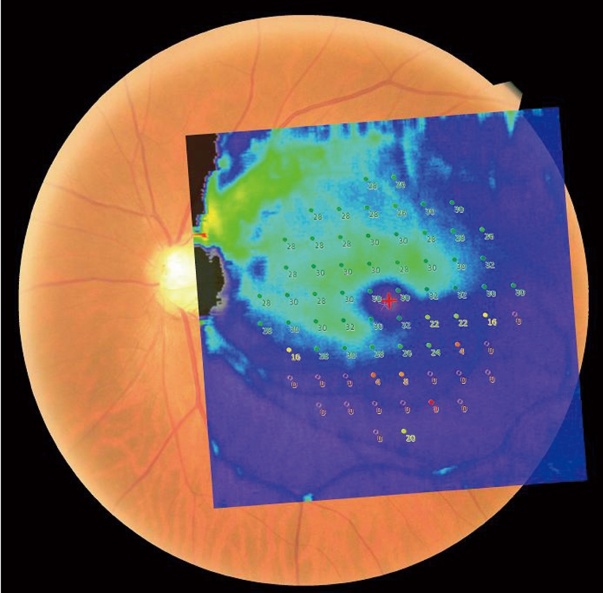
Várias modalidades de OCT podem ser registradas com microperimetria capturada pelo MP-3 da NIDEK.
* Disponível para o modelo Mirante SLO/OCT.
Luigi Sacco Hospital, University of Milan, Italy
Doheny Eye Center, UCLA, USA
Retina Foundation & Eye Research Center, India
Exilaser Clinic, Peru
APAO 2026 – From Pixels to Pathology: The Synergy of DL-Driven OCT and Wide-field Color SLO with Functional Imaging
PAAO 2025 – Unique Capabilities of the NIDEK Mirante
EURETINA 2025
EURETINA 2024
NIDEK’s Mirante Retro mode
APAO 2024
EURETINA 2023
ESCRS 2023
Seeing the pulse of the retina
Emerging role of Mirante
EURETINA 2022
Expert Experience with Multimodal Imaging
Retina Months 2021
Retina Months 2021 *
Mirante Panel Discussion 2020
Multimodal Imaging with the Mirante
Multimodal & Widefield Imaging
Applications of Retro Mode Imaging
The importance of multimodal imaging
WOC 2020
EURETINA 2019
EURETINA 2019 *
FLORetina 2019
Stanislao Rizzo, MD
Professor, Head of Unit of Ophthalmology
Fondazione Policlinico A Gemelli IRCCS & Università Cattolica Sacro Cuore, Italy
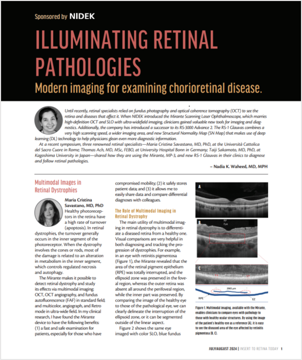
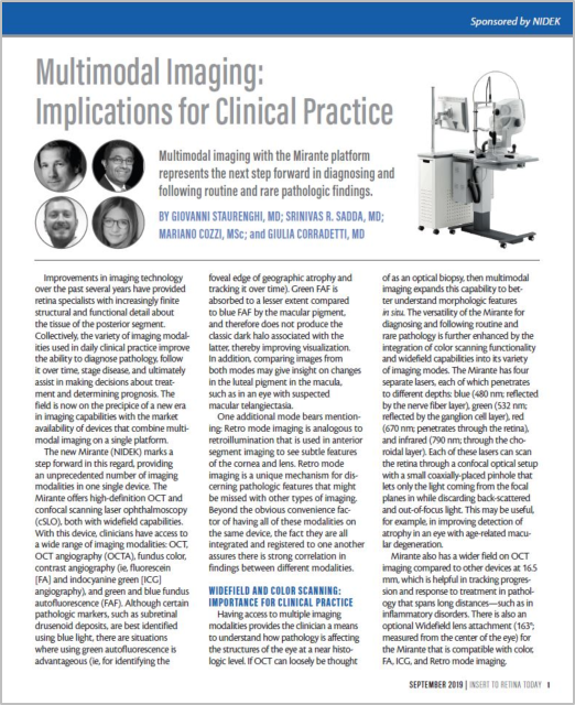
The Mirante multimodal imaging platform integrates multiple clinically significant imaging technologies into a single platform. With this device, clinicians have access to a wide range of imaging modalities including color fundus retinography (SLO), contrast angiography (fluorescein and indocyanine green angiography), green and blue fundus autofluorescence, OCT, OCT angiography (OCTA), and finally the new Retro mode technique. Registration and tracking software allows greater correlation of anatomy, structure and function of the various modalities in this all-in-one device. Additionally, software integration with the NIDEK MP-3 microperimeter can be performed, to correlate (overlay) visual function to structural changes.
The inclusion of ultra wide field imaging allows enhanced evaluation of chorioretinal pathologies involving the periphery, allowing better patient management.
SriniVas R. Sadda, MD
Director, Artificial Intelligence & Imaging Research, Doheny Eye Institute
Professor of Ophthalmology, The University of California – Los Angeles (UCLA), USA
The Mirante platform is a versatile multimodal imaging tool which has proved to be invaluable for diagnosis, staging and management of various retinal diseases in our clinical practice. I have been impressed by the SLO true color fundus photography which provides an accurate color rendition but also offers better contrast and clarity compared with traditional flash color photography. One unique imaging modality of the Mirante device that has been of particular interest to our group is Retro mode imaging, which produces especially high-contrast images with a pseudo-three-dimensional appearance that enhances our ability to detect fine and subtle retinal abnormalities. Notably, the Retro mode and SLO color, as well as other imaging modes (e.g. green and blue fundus autofluorescence, indocyanine angiography, fluorescein angiography) can also be acquired using a wide-angle lens allowing the full extent of the pathology to be evaluated. OCT and OCT angiography are additional Mirante modalities that we have found to be of benefit. Overall, the Mirante is a powerhouse device that allows us to comprehensively assess retinal diseases in our patients.
This product is not cleared by the FDA for distribution in the United States.
Akihiro Ishibazawa, MD, PhD
Manager of Department of Ophthalmology, Japan Red Cross Kitami Hospital, Japan
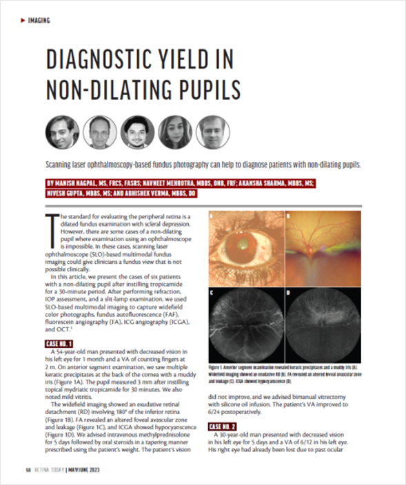
The Mirante multi-color scanning laser ophthalmoscope (SLO) easily captures high-quality images of the posterior-pole and widefield images even in the presence of media opacity. The field of view (FOV) on a single wide image is adequate for documenting the findings of retinal vascular diseases. Changing the fixation target or device angle enables documentation of peripheral retinal breaks and retinal detachment.
Furthermore, macular imaging can be documented in exquisite detail, as can retinal folds due to epiretinal membrane, glaucomatous cupping of the optic disc, and focal retinal nerve fiber defects, etc. As an added benefit, the Mirante performs fluorescein/indocyanine green angiography on the same axis as the color SLO allowing direct correlation of structure to function. The addition of OCT and OCTA allow a thorough analysis of retinal structure and comparison to normative databases. Mirante is a promising new-generation multimodal imaging device for daily use in clinical practice.
Gerardo Garcia-Aguirre, MD
Attending physician, Asociacion para Evitar la Ceguera en Mexico
Clinical professor of Ophthalmology, School of Medicine and Health Sciences, Tecnologico de Monterrey, Mexico
The NIDEK Mirante is a very convenient and practical device for obtaining images of vitreoretinal pathology using different imaging modalities in the same device. It saves both me and the patient the time and hassle of moving between multiple devices. I like the customization capabilities (adjustable scan area and image quality) that I find are beneficial and allow the quick acquisition of images with exceptional resolution in cooperative patients and in patients with poor fixation. Furthermore, the Mirante has some unique, clinically important imaging modalities that are not available in other devices, such as the infrared Retro mode to visualize drusen, and the ultra wide-field infrared SLO to visualize vitreous floaters.
Manish Nagpal, MD
Senior VR Consultant, Retina Foundation, India
Being a laser based SLO device, it allows seamless capturing of great quality images without the requirement of dilation. It is extremely efficient, user friendly, and takes reasonably good images even with dense cataracts.
The ability of Mirante to capture multimodal images allows us to show these to the patients and help counsel them based on their pathologies. It gives us great insight into various retinal pathologies and helps us make accurate treatment decisions.
Moreover, its ability to capture a wide range of images helps us document a lot of images academically, which can be used for teaching as well as sharing on various CME forums to enhance knowledge.
Fabio Patelli, MD
Director, VitreoRetinal service, ASST Santi Paolo e Carlo Hospital, University of Milan
VR consultant, Advalia Vision, Italy
Mirante has all the advantages a physician prefers: high quality images from the SLO, acquisition of wide field images without dilation, increased efficiency for clinical workup by incorporating OCT, OCT-A, Fluorescein Angiography and ICG, fundus autofluorescence and Retro mode all in one device.
Multimodal imaging is now essential for medical retina and Mirante is the first device that incorporates all these characteristics for enhanced diagnostic screening. The high-quality wide field images are fundamental for resident training and patient education.
Shozo Sonoda, MD, PhD
Director, Kagoshima Sonoda Eye Clinic & Plastic Surgery
Visiting Professor, Department of Ophthalmology, Kagoshima University, Japan
The evolution of fundus imaging equipment has been remarkable with the current devices enabling rapid acquisition of high-definition retinal images. While this benefits patients through improved diagnostic outcomes, there is issue of “physicians having to assess an ever-increasing quantity and variety of medical images” and the related possibility of overlooking important details.
Mirante is at the forefront of addressing this issue. For example, as a multimodal device the Mirante acquires high-definition, ultra wide field fundus images that presents more information than conventional fundus cameras. Understanding the principles of this scanning laser ophthalmoscope (SLO) and familiarity with image interpretation enables timely and efficient identification of subtle retinal lesions without overlooking important characteristics. Additionally, the clear high definition images generated by the Mirante facilitate easy patient education thereby encouraging them to participate in treatment.
Nikolle Tan, MD
Consultant Eye Surgeon, Asia Eye Centre, Singapore
The Mirante is a sleek all-in-one machine that makes multimodal imaging for retinal diseases a breeze. With its high definition confocal SLO, OCT, and OCT-A functions, the Mirante is a reliable and consistent workhorse that should be the foundation of any busy Retina practice.
It also comes with an exceptional number of other valuable functions, including ultra-wide-field imaging options, fundus autofluorescence and Retro mode. I find the ultra-wide-field imaging capability of the Mirante particularly indispensable, as in addition to ultra-wide-field color fundus photography, the system also allows for ultra-wide-field simultaneous FA and ICG.
The learning curve for using the Mirante is not steep, and together with Mirante’s excellent technical support team who also spares no effort in training users of this machine, it is easy for anyone, even a junior technician, to acquire consistently quality images.